
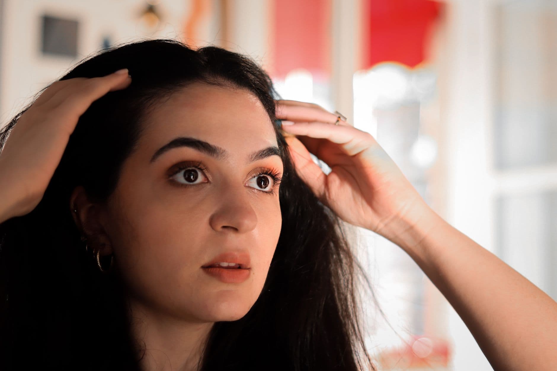
For women living and working in fast-paced corporate and residential neighbourhoods across Pune—such as Kalyani Nagar, Viman Nagar, and Koregaon Park—noticing a widening central parting or gradual loss of ponytail volume is an increasingly common concern. Demanding work hours, chronic urban stress, environmental pollution, and hormonal shifts frequently lead people to assume that hair shedding is simply an unavoidable reaction to modern life. However, sustained thinning is distinct from ordinary daily hair shedding, representing a specific biological change in follicular health.
Female pattern hair loss (FPHL) behaves quite differently from male pattern baldness. While men often present with frontotemporal hairline recession, women typically experience a diffuse reduction in hair density over the crown and frontal scalp while retaining their frontal hairline fringe (Female pattern hair loss). Because this onset is gradual, many individuals spend months relying on commercial anti-hairfall shampoos, salon spa rituals, or unverified home oils, none of which address progressive follicular changes.
Addressing chronic thinning effectively requires looking beneath surface symptoms. Consulting a medical dermatologist provides access to clinical scalp evaluations and objective diagnostic tools capable of pinpointing the underlying hormonal, genetic, and environmental factors driving hair loss, enabling targeted medical management before follicular density is compromised.
Recognising the Clinical Signs: How Female Pattern Thinning Manifests
Dermatologists categorise the progression of female pattern hair loss using structured clinical classifications rather than subjective observation. The most widely recognised framework is Ludwig’s classification, which outlines three progressive stages of thinning (Female pattern hair loss):
- Stage I: Characterised by perceptible thinning of hair over the anterior crown with minimal widening or rarefaction of the part width (Female pattern hair loss).
- Stage II: Marked by more pronounced rarefaction of the crown hair, accompanied by an increased proportion of noticeably shorter and thinner shafts (Female pattern hair loss).
- Stage III: Characterised by near-total, diffuse baldness across the crown, though an intact frontal hairline fringe is typically preserved (Female pattern hair loss).
In clinical practice, dermatologists also recognise alternative presentation patterns. Olsen emphasized frontal accentuation, known as the 'Christmas tree' pattern of hair loss in female pattern hair loss (Female pattern hair loss). Other frameworks, such as the Savin hair density scale and the Basic and Specific (BASP) classification system, categorize presentations based on observed patterns of hair loss (Female pattern hair loss).
A key challenge in female hair loss is that standard observational scales like the Ludwig and Sinclair systems rely on assessing visible hair thinning on the crown, which is the reason they often fail to detect early-stage female pattern hair loss (Utility of trichoscopy to diagnose early female pattern hair loss i…). Microscopic alterations often emerge within follicles well before crown rarefaction becomes conspicuous during an unaided visual examination.
Urban Pune Triggers and the Metabolic Link: Hormones, PCOS, and Skin Health
When investigating hair thinning in urban centres like Pune, dermatologists evaluate internal systemic drivers alongside external environmental triggers. Chief among metabolic factors is hyperandrogenism, often connected to Polycystic Ovary Syndrome (PCOS) (Female pattern hair loss, Polycystic ovary syndrome). PCOS is diagnosed when a patient meets at least two of three clinical criteria: menstrual disturbance, clinical or biochemical hyperandrogenism, and ultrasound-detected polycystic ovaries (Polycystic ovary syndrome).
The physiological link between ovarian dysfunction and hair thinning is well established. In a clinical study of 89 women presenting with hair loss, 67% showed ultrasound evidence of polycystic ovaries compared to 27% in the control group, and 21% exhibited significant hirsutism compared to 4% of controls (Female pattern hair loss). The condition is thought to develop when the ovaries overproduce androgens such as testosterone as a result of elevated luteinising hormone secretion from the pituitary gland, alongside peripheral insulin resistance (Polycystic ovary syndrome).
This hormonal environment does not affect the scalp in isolation. In polycystic ovary syndrome, elevated androgen levels can lead to female pattern or male pattern hair loss alongside cutaneous manifestations such as persistent acne, oily skin (seborrhoea), acanthosis nigricans, and hirsutism—excess hair growth on the face, chest, abdomen, thumbs, or toes (Polycystic ovary syndrome). Consequently, addressing female pattern thinning often forms part of an integrated dermatological strategy that incorporates medical acne treatment, procedures for acne scars, and laser hair reduction for unwanted facial hair.
In addition to metabolic shifts, residents in Pune encounter local environmental stressors. Hard water supplied in various urban pockets contains mineral salts that can deposit along the hair shaft, contributing to moisture loss, strand brittleness, and scalp dryness. When paired with high-stress corporate schedules, dietary gaps such as low iron or vitamin deficiencies, and fluctuating seasonal humidity, the scalp can develop low-grade inflammation that compounds hormonal thinning.
The Dermatological Diagnostic Pathway: Trichoscopy and Scalp Health Mapping
To achieve an accurate assessment, MD dermatologists evaluate microscopic follicular structures using trichoscopy and dermoscopy (Utility of trichoscopy to diagnose early female pattern hair loss i…). Microscopic changes recorded with dermoscopy are helpful in establishing a diagnosis of female pattern hair loss even in early disease, avoiding the diagnostic delays common with macroscopic visual scales (Utility of trichoscopy to diagnose early female pattern hair loss i…).
Under healthy conditions, normal scalp architecture features follicular units containing 1 to 4 terminal hairs with 1 to 2 vellus hairs (Utility of trichoscopy to diagnose early female pattern hair loss i…). In female pattern hair loss, this balance shifts markedly: the frontal scalp displays an increased proportion of single-hair follicular units and miniaturised thin hairs (Utility of trichoscopy to diagnose early female pattern hair loss i…). Dermoscopy also readily identifies anisotrichosis—the variation in hair shaft diameters that reflects progressive follicular miniaturisation (Female pattern hair loss, Utility of trichoscopy to diagnose early female pattern hair loss i…).
Major videodermoscopy criteria published by Rakowska et al. provide objective parameters for diagnosing female pattern hair loss (Utility of trichoscopy to diagnose early female pattern hair loss i…):
- More than 4 yellow dots across four images in the frontal area (Utility of trichoscopy to diagnose early female pattern hair loss i…).
- Lower average hair thickness in the frontal area compared with the occiput area (Utility of trichoscopy to diagnose early female pattern hair loss i…).
- More than 10% thin hairs measuring less than 0.03 mm in diameter in the frontal area (Utility of trichoscopy to diagnose early female pattern hair loss i…).
Additional characteristic dermatoscopic features include a brown peripilar halo at the follicular ostium, scalp pigmentation, and empty follicles in post-menopausal women (Female pattern hair loss). In a comparative study evaluating early female pattern hair loss, the peripilar sign was noted in 18.7% of cases and yellow dots in 7.4% of cases, highlighting that while these markers are specific, their occurrence can be less frequent in the earliest stages (Utility of trichoscopy to diagnose early female pattern hair loss i…).
Clinical research demonstrates that microscopic changes recorded with an accessible USB dermoscope can reliably diagnose early female pattern hair loss (disease duration under six months and part width under 1 cm) by identifying anisotrichosis, vellus-like hairs, and single-hair units in the frontal scalp compared to the occipital region (Utility of trichoscopy to diagnose early female pattern hair loss i…). This diagnostic precision ensures that medical management begins on the basis of verified follicular criteria.
Clinical Interventions and Regenerative Procedures: Medical Care and PRP
Managing female pattern thinning requires structured clinical intervention. Because female pattern hair loss is progressive in nature, long-term treatment under medical supervision is mandated to achieve a sustained therapeutic effect (Female pattern hair loss).
Medical Stabilisation
Topical minoxidil solution remains the established first-line medical therapy for female pattern hair loss (Female pattern hair loss). In cases where hyperandrogenism or hormonal imbalances are identified, dermatologists may introduce second-line medical therapies, including anti-androgens such as cyproterone acetate, spironolactone, or flutamide (Female pattern hair loss). While anti-androgenic options like spironolactone can help reduce persistent acne and excess body hair over time in patients with PCOS, clinical evidence indicates they are less effective for hair loss, highlighting the importance of doctor-guided treatment selection (Polycystic ovary syndrome).
Platelet-Rich Plasma (PRP) Therapy
Autologous Platelet-Rich Plasma (PRP) has been extensively evaluated in clinical studies for the treatment of female pattern hair loss (Platelet-Rich Plasma in Female Androgenic Alopecia: A Comprehensive…). PRP is prepared by processing the patient's peripheral blood to concentrate platelets containing major regenerative growth factors (Platelet-Rich Plasma in Female Androgenic Alopecia: A Comprehensive…). Published data highlight that PRP contains key biological molecules, including (Platelet-Rich Plasma in Female Androgenic Alopecia: A Comprehensive…):
- Vascular Endothelial Growth Factor (VEGF) (Platelet-Rich Plasma in Female Androgenic Alopecia: A Comprehensive…).
- Platelet-Derived Growth Factor (PDGF) (Platelet-Rich Plasma in Female Androgenic Alopecia: A Comprehensive…).
- Basic Fibroblast Growth Factor (bFGF) (Platelet-Rich Plasma in Female Androgenic Alopecia: A Comprehensive…).
- Epidermal Growth Factor (EGF) (Platelet-Rich Plasma in Female Androgenic Alopecia: A Comprehensive…).
- Transforming Growth Factor-β (TGF-β) (Platelet-Rich Plasma in Female Androgenic Alopecia: A Comprehensive…).
- Insulin-Like Growth Factor-1 (IGF-1) (Platelet-Rich Plasma in Female Androgenic Alopecia: A Comprehensive…).
The efficacy of PRP in treating female pattern hair loss has been confirmed by comprehensive clinical research. In a systematic review and meta-analysis of 42 studies comprising 1,569 cases (including 776 female participants presenting mostly with type I to III FPHL on the Ludwig scale or grades 1 to 5 on the Sinclair score system), PRP demonstrated positive efficacy in increasing hair density compared to control groups (odds ratio 1.61, 95% CI 0.52–2.70) (Platelet-Rich Plasma in Female Androgenic Alopecia: A Comprehensive…). Furthermore, when compared directly to baseline measurements using a random-effects model, PRP demonstrated significant improvements in hair density (odds ratio 1.11, 95% CI 0.86–1.37) (Platelet-Rich Plasma in Female Androgenic Alopecia: A Comprehensive…).
Realistic Expectations and Safety
Clinical treatments require realistic timelines and transparent discussions. PRP is designed to support and stimulate existing follicular structures rather than reviving areas where follicular tissue is absent. Documented adverse effects in clinical trials are mild and resolve spontaneously: patients may experience mild headache, scalp sensitivity, or bruising and mild pain at injection sites after 48 to 72 hours, which resolve spontaneously by the fourth or fifth post-procedure day (Platelet-Rich Plasma in Female Androgenic Alopecia: A Comprehensive…).
Formulating a Personalised Care Strategy in Kalyani Nagar
Every scalp is biologically unique, meaning that generalised commercial packages cannot replace an evaluation tailored to an individual’s medical profile. In an urban setting like Pune, an effective hair preservation plan must account for hair calibre, familial predisposition, metabolic indicators like insulin resistance, and daily lifestyle stresses.
A comprehensive strategy also addresses scalp hygiene and environmental management. Pune's hard water and warm, humid periods can cause mineral deposits to combine with sebum, fostering an environment where inflammatory microbes thrive. Managing this involves using gentle, non-stripping scalp cleansers to remove residue and dead skin cells without depleting essential barrier lipids, protecting vulnerable miniaturising shafts from breakage.
Furthermore, because female pattern thinning frequently overlaps with conditions like PCOS, an effective dermatological strategy should harmonize hair restoration with broader skin health goals. Women managing hair thinning alongside hormonal acne, post-inflammatory hyperpigmentation, or unwanted facial hirsutism benefit from a coordinated clinical roadmap. Integrating medical acne care, acne scar treatments, laser toning for pigmentation, and laser hair reduction alongside follicular therapies ensures that aesthetic and dermatological concerns are addressed cohesively.
Taking a Clinical Approach to Hair Restoration in Pune
Female hair thinning is a progressive medical condition driven by follicular miniaturisation, metabolic influences, and hormonal factors (Female pattern hair loss, Polycystic ovary syndrome). Relying on cosmetic cover-ups or waiting for widening part lines to resolve spontaneously allows miniaturisation to advance unchecked, making early professional intervention critical. Microscopic dermoscopy and clinical therapies offer a scientific pathway to diagnose follicular changes and support hair density (Utility of trichoscopy to diagnose early female pattern hair loss i…, Platelet-Rich Plasma in Female Androgenic Alopecia: A Comprehensive…).
At Skin Essence in Kalyani Nagar, Pune, clinical hair loss evaluations and medical therapies are led by Dr. Daksha Patel, MBBS, MD (Skin & VD), a dermatologist and cosmetologist. Rather than relying on rigid protocols, each treatment plan—from diagnostic mapping and medical hair loss management to PRP hair treatment and medical dermatology—is individually formulated around the patient's specific scalp health, skin type, and lifestyle. For individuals seeking clarity on diffuse shedding or part-line widening, scheduling a clinical consultation with an MD dermatologist is the essential first step toward sustainable hair health.
Frequently asked questions
How does female pattern hair loss differ from male balding?
Male pattern baldness typically involves frontotemporal hairline recession, whereas female pattern hair loss causes diffuse thinning across the crown and frontal scalp while usually preserving the frontal hairline fringe.
Why is female hair thinning linked to conditions like PCOS?
Polycystic ovary syndrome involves elevated androgen levels and peripheral insulin resistance, which can drive follicular miniaturisation on the scalp alongside symptoms like persistent acne and hirsutism.
How do dermatologists diagnose early female pattern hair loss?
Dermatologists use dermoscopy and trichoscopy to identify microscopic changes such as hair shaft diameter variation (anisotrichosis), an increase in single-hair units, and yellow dots before thinning is clearly visible to the naked eye.
What role does PRP play in managing female hair thinning?
Platelet-Rich Plasma (PRP) uses concentrated autologous platelets containing growth factors like VEGF and PDGF to support and stimulate existing hair follicles, increasing hair density over time.