
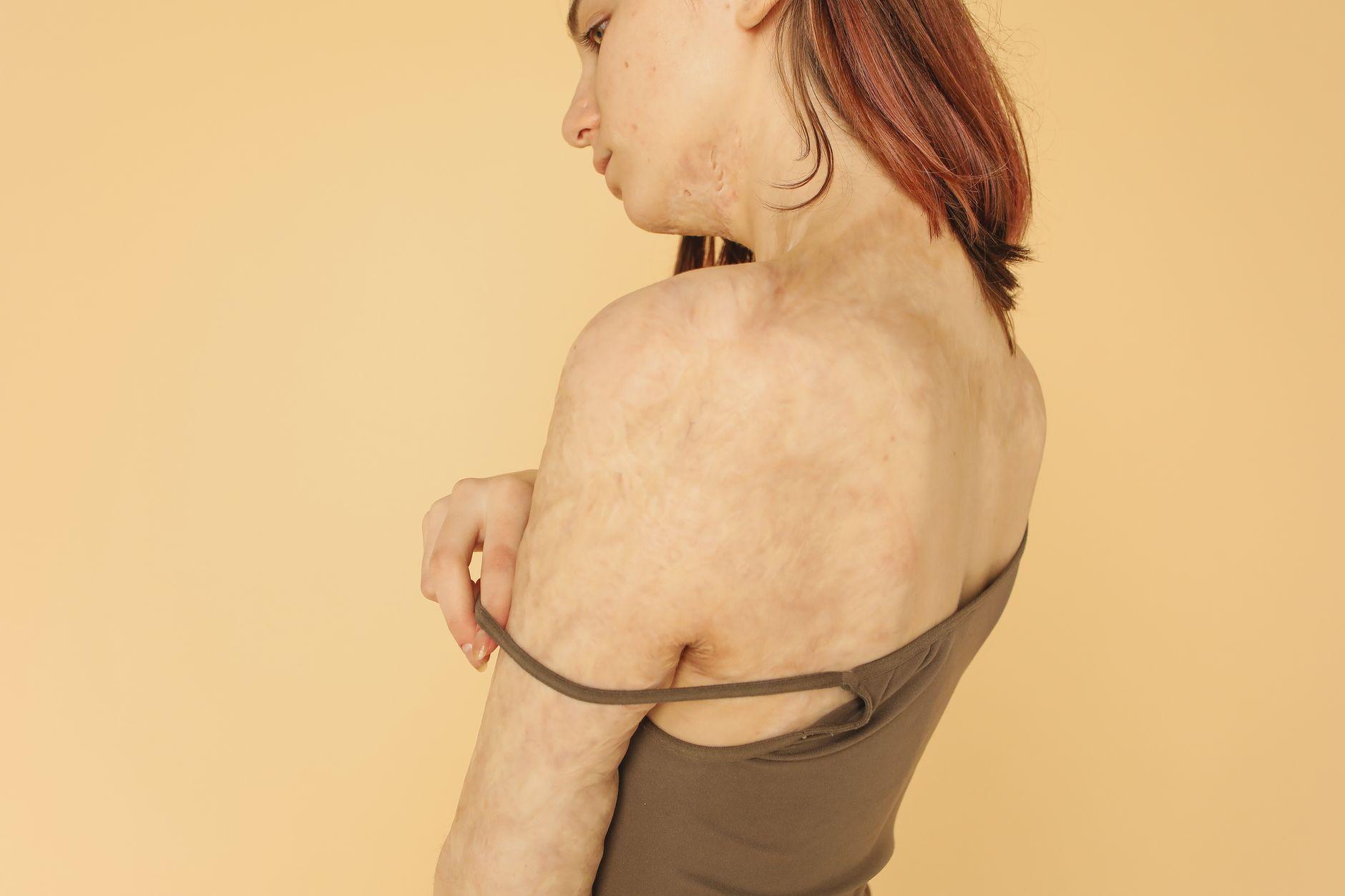
For many people in Pune, severe breakouts leave behind unexpected marks. Most discussions focus on pitted or depressed acne marks. Yet, breakouts on the jawline, neck, and chest often leave firm, elevated lumps. These raised areas are hypertrophic acne scars. They show increased firmness and altered colour restricted strictly to the site of the original injury (An Updated Review of Hypertrophic Scarring).
Treating raised scars demands a very different strategy than treating shallow pits. Harsh scrubs and strong chemical exfoliants cause irritation. For melanin-rich Indian skin tones, this excess friction frequently triggers post-inflammatory hyperpigmentation. Pune has warm weather and strong sunlight for much of the year. In this climate, protecting healing skin from pigment changes is just as vital as flattening the scar tissue.
Smoothing thickened skin requires regulating collagen production and softening firm fibrous tissue. It also requires calming local inflammation. A clinical consultation at a medical dermatology centre in Kalyani Nagar, Pune helps patients identify their marks accurately. This assessment ensures each person receives a safe, step-by-step scar remodelling plan.
The Biology of Elevated Blemishes: Why Excess Collagen Forms
Raised acne scars develop when normal wound healing goes off track after deep breakouts. During normal tissue repair, new structural tissue forms and then balances out. Pathologic scarring happens when fibroblasts multiply excessively and turn into myofibroblasts (Hydrogel Loaded with Components for Therapeutic Applications in Hyp…). This creates an imbalance where extracellular matrix deposition outpaces matrix breakdown (Hydrogel Loaded with Components for Therapeutic Applications in Hyp…).
At a microscopic level, structural buildup happens because normal breakdown enzymes decline. Hypertrophic and keloidal acne scars develop from reduced collagenase activity paired with exuberant collagen deposition (Evidence-based Surgical Management of Post-acne Scarring in Skin of…). During the remodelling stage, matrix metalloproteinases MMP1 and MMP7 drop (An Updated Review of Hypertrophic Scarring). This drop slows the normal clearance of structural proteins like collagen type I, collagen type III, and fibronectin (An Updated Review of Hypertrophic Scarring). At the same time, tissue inhibitors of metalloproteinases TIMP1 and TIMP2 block remaining enzyme action (An Updated Review of Hypertrophic Scarring). This process locks dense tissue in place.
Ongoing dermal inflammation fuels this cellular overactivity. Specialised immune cells called M2 macrophages release growth signals like TGF-beta and PDGF-CC (The Roles of Inflammation in Keloid and Hypertrophic Scars). These signals prompt local fibroblasts to change into contractile myofibroblasts (The Roles of Inflammation in Keloid and Hypertrophic Scars). As a result, abnormal collagen continues to build up (The Roles of Inflammation in Keloid and Hypertrophic Scars).
Physical movement also shapes how marks heal. Hypertrophic scars often appear in high-tension regions like the shoulders, neck, and prosternum (An Updated Review of Hypertrophic Scarring). Constant skin tension on areas like the anterior chest promotes fibrosis through a mechanotransduction pathway driven by focal adhesion kinase (The Roles of Inflammation in Keloid and Hypertrophic Scars). High mechanical skin tension stretches cells and tissue, activating cascades such as FAK/ERK and YAP/TAZ (Hydrogel Loaded with Components for Therapeutic Applications in Hyp…). These pathways cause fibroblasts to multiply and produce excess matrix (Hydrogel Loaded with Components for Therapeutic Applications in Hyp…). In areas subject to daily stretching, acne blemishes frequently heal as raised, firm marks.
Hypertrophic Acne Scars Versus Keloids: Identifying Your Lesions
Raised blemishes along the jawline, lower face, and torso can look similar at first glance. However, their clinical behaviour and long-term outlook vary widely. A qualified dermatologist must evaluate whether a lesion is a hypertrophic scar or a keloid before starting treatment.
- Hypertrophic Scars: These marks stay confined within the margins of the initial wound (Standard guidelines of care: Keloids and hypertrophic scars). They are typically softer lesions with normal skin colouration (Hydrogel Loaded with Components for Therapeutic Applications in Hyp…). Over time, they may undergo partial spontaneous resolution without invading surrounding skin (Standard guidelines of care: Keloids and hypertrophic scars, Hydrogel Loaded with Components for Therapeutic Applications in Hyp…). Under a microscope, they show organized bundles of type III collagen (Hydrogel Loaded with Components for Therapeutic Applications in Hyp…).
- Keloidal Lesions: Unlike hypertrophic scars that remain within the initial wound boundaries, keloids extend beyond the margins of the original wound (Standard guidelines of care: Keloids and hypertrophic scars). They appear clinically as firm, raised, and pigmented scars that rarely regress spontaneously and tend to recur after excision (Standard guidelines of care: Keloids and hypertrophic scars, Hydrogel Loaded with Components for Therapeutic Applications in Hyp…). Histologically, they feature thick, disorganized bundles of type I and type III collagen (Hydrogel Loaded with Components for Therapeutic Applications in Hyp…).
For Indian skin tones, this clinical distinction is essential. Treating an active keloid with unsuitable methods can irritate the tissue and cause it to expand further. An evaluation by an MD dermatologist confirms lesion borders, firmness, and activity. This ensures the patient receives an appropriate and tailored management plan.
Targeted In-Clinic Dermal Procedures for Tissue Softening
Over-the-counter creams rarely reach deep enough to remodel dense fibrous tissue. In clinical dermatology, targeted intradermal procedures deliver active solutions directly into the scar. Intradermal administration of a prescription anti-inflammatory medicine reduces excess scarring by calming inflammatory activity across myeloid and lymphoid cells (The Roles of Inflammation in Keloid and Hypertrophic Scars). Your dermatologist will advise whether this is suitable for you.
Accurate depth of placement is vital during these clinical sessions. Practice guidelines state that the prescription anti-inflammatory agent must be introduced into the mid-dermis (Standard guidelines of care: Keloids and hypertrophic scars). If the solution is placed too superficially, it can cause irreversible epidermal atrophy (Standard guidelines of care: Keloids and hypertrophic scars). Higher strengths can also lead to higher rates of skin thinning and visible telangiectasias (An Updated Review of Hypertrophic Scarring). Sessions are planned carefully over time, and your dermatologist explains how any prescribed medicine is used.
To improve results and reduce unwanted skin changes, dermatologists often combine active agents. Research demonstrates that pairing a prescription anti-inflammatory medicine with a complementary clinical compound achieves a 92% average reduction in lesion size, compared to 73% with the anti-inflammatory alone (Standard guidelines of care: Keloids and hypertrophic scars). This combined method also produces greater reductions in scar height and lower rates of skin thinning, telangiectasias, and hypopigmentation (An Updated Review of Hypertrophic Scarring). Known side effects of the complementary compound include pain, hyperpigmentation, and tissue sloughing (Standard guidelines of care: Keloids and hypertrophic scars). Your dermatologist will advise whether this is suitable for you.
Laser Care and Energy-Based Devices for Melanin-Rich Skin
Energy-based treatments support dermal procedures by remodelling collagen architecture and calming microvascular activity. In Fitzpatrick skin types IV and V, epidermal melanin readily absorbs light energy. Because of this, lasers must be calibrated carefully to prevent post-inflammatory hyperpigmentation.
For raised acne scars with lingering redness, pulsed dye laser (PDL) holds a Level of Evidence I and Strength of Recommendation A rating (Evidence-based Surgical Management of Post-acne Scarring in Skin of…). Clinical guidelines identify pulsed dye vascular lasers as the laser treatment of choice for hypertrophic acne scars (An Updated Review of Hypertrophic Scarring). In dense scar tissue, pulsed dye lasers help decrease TGF-beta1 induction and upregulate matrix metalloproteinase expression (Standard guidelines of care: Keloids and hypertrophic scars). These lasers can also assist the clinical delivery of localized therapeutic agents into the scar tissue (An Updated Review of Hypertrophic Scarring).
When using fractional energy systems to soften scars, settings must match melanin-rich skin:
- Non-Ablative Fractional Settings: For skin types IV and V, non-ablative fractional devices like 1550 nm erbium-doped lasers use pulse energies between 30 and 70 mJ (Evidence-based Surgical Management of Post-acne Scarring in Skin of…). These settings remodel deeper collagen while keeping the skin barrier intact.
- Conservative Fluence and Density: Lower energy fluences are advised in darker complexions to minimise post-inflammatory hyperpigmentation (Evidence-based Surgical Management of Post-acne Scarring in Skin of…).
- Avoiding Unsuitable Monotherapy: Carbon dioxide laser monotherapy carries a recurrence rate reaching 90% and is not recommended for raised scars (Standard guidelines of care: Keloids and hypertrophic scars). In addition, post-inflammatory hyperpigmentation occurs in 6.4% to 92.3% of skin type IV and V patients following fractional carbon dioxide treatments, with higher fluences and densities linked to greater pigment risk (Evidence-based Surgical Management of Post-acne Scarring in Skin of…).
Using conservative energy levels and avoiding areas with active surface infections allows clinicians to remodel firm scar tissue safely without causing pigment issues.
Daily Skin Care: Active Breakout Control and Pigment Defence
Flattening established marks is only part of scar care. Long-term skin health requires stopping new breakouts, protecting healing tissue from UV rays, and reducing surface friction.
Halting Active Acne Breakouts
Every new inflamed cyst can restart abnormal wound repair and generate fresh fibrous tissue. Structured acne treatment is therefore an essential clinical priority. Calming deep follicular inflammation prevents the release of profibrotic signals before they trigger myofibroblasts. Once active breakouts settle, acne scar treatment proceeds with far less risk of new scar formation.
Minimising Jawline Friction and Follicular Irritation
Along the jawline and neck, recurring ingrown hairs, folliculitis, and daily shaving create mechanical friction. This friction maintains local inflammation and high tissue stress. For suitable candidates, laser hair reduction planned for Indian skin tones removes the irritation of coarse hair regrowth. Clearing chronic beard-area irritation relieves mechanical strain and reduces triggers that promote raised scars.
Daily Photoprotection and Gentle Skin Handling
Healing tissue contains reactive pigment-producing cells that respond strongly to ultraviolet light. In Pune's sunny climate, unshielded sun exposure stimulates excess melanin, turning pink marks dark brown. Applying broad-spectrum sunscreen every morning shields remodelling skin from UV-induced darkening.
Patients should also avoid picking at bumps or scrubbing the skin with stiff sponges. Rough handling increases skin tension and triggers cellular mechanotransduction pathways, prompting fibroblasts to create more matrix. Gentle daily washing combined with medical guidance supports steady, smooth healing.
Personalised Care for Thickened Marks in Pune
Managing raised acne scars requires an understanding of wound biology, collagen turnover, and melanin safety. Because these marks stem from excess tissue rather than tissue loss, standard resurfacing methods are not enough on their own. Successful management combines targeted dermal procedures to soften fibrous bundles, conservative laser care to guide collagen remodelling, and consistent control of active blemishes.
For individuals seeking guidance in Kalyani Nagar, Pune, an in-person assessment allows an MD dermatologist to examine lesion margins, scar firmness, and skin reactivity. If you are dealing with firm, raised marks on your jawline or chest, call the clinic, WhatsApp the clinic, or send an enquiry to arrange a clinical evaluation.
Frequently asked questions
What causes hypertrophic acne scars to form?
They develop when wound repair after deep breakouts leads to excess collagen deposition and decreased natural breakdown enzymes. Dermal inflammation and mechanical skin tension also prompt fibroblasts to produce dense fibrous matrix.
How are hypertrophic acne scars different from keloids?
Hypertrophic scars stay strictly within the boundaries of the original breakout and may partially regress over time. Keloids extend beyond the initial wound borders, rarely regress on their own, and tend to recur.
Which laser treatments are used for raised acne scars on Indian skin tones?
Pulsed dye vascular lasers are commonly chosen to address microvascular activity and help remodel collagen in raised scars. For deeper remodelling in melanin-rich skin, non-ablative fractional devices at conservative fluences are preferred over high-risk ablative monotherapies.
Why is active acne control important while treating raised scars?
New inflamed blemishes release profibrotic signals that trigger myofibroblasts and abnormal wound repair. Halting active breakouts prevents fresh fibrous tissue and raised marks from developing.
This information is general and not a substitute for a consultation. Results vary from person to person.